reSee.it - Tweets Saved By @DrJohnB2

Saved - June 30, 2024 at 5:18 AM
reSee.it AI Summary
White blood clots found by embalmers, possibly related to COVID-19 vaccinations, have been studied by Richard Hirschman. The clots contain proteins such as fibrinogen, hemoglobin, fibronectin, and von Willebrand factor. The composition and color of the clots can indicate underlying pathologies. White clots are less common and contain more platelets and collagen/calcification. Abnormal fibrin polymerization and the presence of immunoglobulin heavy constant gamma 1 protein and myeloperoxidase may contribute to the formation of these clots. Neutrophil extracellular traps (NETs) are also likely involved.

@DrJohnB2 - Dr John B.

(1/n) What are these generally white blood clots found by various embalmers, probably mainly related to COVID-19 vaccinations? Embalmer Richard Hirschman (@r_hirschman) has now published some interesting findings about these clots (https://x.com/r_hirschman/status/1800562643888931174). Here is a summary and discussion. These are the proteins that were found:

@r_hirschman - Richard Hirschman

Here's a list of the top 21 proteins found in the strange fibrous clots. There were over 500 proteins found. I am not a doctor or scientist therefore I don't fully understand what this is saying, but I am hoping that those who can interpret the information might learn from it. One thing that I was told was thrombin wasn't detected. Here is also the results of the amino acids detected. Hopefully this will help those trying to figure out what this is and hopefully help humanity. I think these tests should be replicated just like the ICP-MS that Mike Adams did. Most people like myself will not understand what these tests are saying, most of us are not trained to understand this, but there are some people out there that might. Seek the truth and God 🙏 Be kind 😇 Try to do what is right ✅️

@DrJohnB2 - Dr John B.

(2/n) The most abundant protein is fibrinogen (fibrinogen chains alpha, beta & gamma) (making 56% of total clot protein abundance). Followed by hemoglobin (subunit beta and alpha). Fibronectin is an important protein for cell adhesion (and in a clot is normally covalently cross-linked to fibin by Factor XIII). Fibrinogen chains are the main constituents of blood clots, and the presence of relatively large amounts of fibronectin is typical. See, for example, the study of plasma clots by Michał Ząbczyk et al. [1]: [1] https://www.sciencedirect.com/science/article/abs/pii/S1874391919302593?casa_token=t_uXsXq6Gc0AAAAA:UGLeoVazv33jKhyWucjgvS6dg2AVn3f0Vktxp-TkF43klqHlLQCuVGzT4lCRVSK_pMiNqXbVlg

Plasma fibrin clot proteomics in healthy subjects: Relation to clot permeability and lysis time Little is known about fibrin clot composition in relation to its structure and lysability. We investigated plasma clots protein composition and its as… sciencedirect.com

@DrJohnB2 - Dr John B.

(3/n) The hemoglobin proteins (beta & alpha subunits) indicate the presence of erythrocytes or the hemoglobin of the erythrocytes (free hemoglobin, Hb). Hb is known to bind to von Willebrand Factor (VWF), inducing the formation of platelet aggregates on fibrinogen-rich surfaces [1]. The amount of erythrocytes/Hb present in clots varies. Choi et al [2] showed that the more erythrocytes there are in a clot, the more red it is: [1] https://ashpublications.org/blood/article/126/20/2262/103848/Free-hemoglobin-a-boost-to-platelet-thrombi [2] https://www.ahajournals.org/doi/full/10.1161/STROKEAHA.117.019138

@DrJohnB2 - Dr John B.

(4/n) In terms of clot color, the relative abundance of erythrocytes and fibrin is related to the color of the clot and the ratio is related to the physical properties of the clot (more erythrocytes in the clot: more soft & friable clot) [1]: [1] https://neuronewsinternational.com/the-importance-of-clot-properties-in-endovascular-stroke-therapy

The importance of clot properties in endovascular stroke therapy - NeuroNews International By Tommy Andersson Recently no less than five randomised controlled trials showed superiority for intra-arterial treatment added to intravenous thrombolysis (IVT) of large artery stroke in the anterior circulation as compared to stand-alone IVT1–5. This obvious breakthrough is more than welcomed, but it is also clear from these studies that challenges remain, not least for […] neuronewsinternational.com

@DrJohnB2 - Dr John B.

(5/n) Sgreccia et al [1] concluded that the color of the clot is related to its composition and could be a „warning light for underlying pathologies“. White clots are less common and indicate an "atypical" aetiology: [1] https://jnis.bmj.com/content/11/12/1197

Association between acute ischemic stroke etiology and macroscopic aspect of retrieved clots: is a clot’s color a warning light for underlying pathologies? Background Few case reports have considered the chromatic aspect of retrieved clots and the possible association with their underlying etiology. Objective The aim of our study was to analyze the frequency of the TOAST ischemic stroke typical (atrial fibrillation, dissection, atheroma) and atypical (infective endocarditis, cancer-related, valve-related thrombi) etiologies depending on the chromatic aspect of retrieved clots. Methods A total of 255 anonymized and standardized clot photos of consecutive patients treated by mechanical thrombectomy for acute ischemic stroke were included. A double-blind evaluation was performed by two senior interventional neuroradiologists, who classified the visual aspects of the clots into two main patterns: red/black or white. Main patient characteristics, distribution of underlying stroke etiologies, and outcomes were compared between the two study groups. Results The inter-reader agreement for clot colors was excellent (k=0.78). Two hundred and thirty-three patients were classified as having red/black clots and 22 as having white clots. A statistically significant association (p=0.001) between atypical etiologies and white clots was observed. Conclusions White clots were significantly associated with atypical etiologies in this cohort,in particular, with infectious endocarditis. jnis.bmj.com

@DrJohnB2 - Dr John B.

(6/n) In a study published in 2021, Mereuta et al [1] examined white clots, as they "represent a less common entity and their histological composition is less known". They highlighted that these white clots „account for a small subset of thrombi and are often perplexing to the clinician regarding the etiology“. The study found that white clots were less common than red clots (14% of the 760 clots studied) and contained significantly more platelets and collagen/calcification and less erythrocytes and white blood cells than red clots. (White clots also contained a significant amount of von Willebrand factor (vWF). vWF was also found in Richard Hirschman's analysis). [1] https://www.sciencedirect.com/science/article/pii/S1052305721005322

Characterization of the ‘White’ Appearing Clots that Cause Acute Ischemic Stroke Most clots retrieved from patients with acute ischemic stroke are ‘red’ in color. ‘White’ clots represent a less common entity and their histological … sciencedirect.com

@DrJohnB2 - Dr John B.

(7/n) In my opinion, the discovery of "fibrinaloid" microclots by Pretorius & Kell [1] is very important in explaining the white clots observed by embalmers. As highlighted in a recent excellent review by Kell et al [2], the polymerisation of fibrinogen to fibrin in clots can occur in different forms on a spectrum. One end of the spectrum represents the normal fibrin polymerisation process in which "normal" (α-helix-rich) fibrin is formed. The other end of the spectrum is characterised by abnormal fibrin polymerisation, resulting in a crossed-β-sheet amyloid 'fibrinaloid' form: [1] https://pubmed.ncbi.nlm.nih.gov/27605168/ [2] https://www.mdpi.com/2227-9059/12/4/891

Acute induction of anomalous and amyloidogenic blood clotting by molecular amplification of highly substoichiometric levels of bacterial lipopolysaccharide - PubMed It is well known that a variety of inflammatory diseases are accompanied by hypercoagulability, and a number of more-or-less longer-term signalling pathways have been shown to be involved. In recent work, we have suggested a direct and primary role for bacterial lipopolysaccharide (LPS) in this hype … pubmed.ncbi.nlm.nih.gov
Fibrinaloid Microclots and Atrial Fibrillation Atrial fibrillation (AF) is a comorbidity of a variety of other chronic, inflammatory diseases for which fibrinaloid microclots are a known accompaniment (and in some cases, a cause, with a mechanistic basis). Clots are, of course, a well-known consequence of atrial fibrillation. We here ask the question whether the fibrinaloid microclots seen in plasma or serum may in fact also be a cause of (or contributor to) the development of AF. We consider known ‘risk factors’ for AF, and in particular, exogenous stimuli such as infection and air pollution by particulates, both of which are known to cause AF. The external accompaniments of both bacterial (lipopolysaccharide and lipoteichoic acids) and viral (SARS-CoV-2 spike protein) infections are known to stimulate fibrinaloid microclots when added in vitro, and fibrinaloid microclots, as with other amyloid proteins, can be cytotoxic, both by inducing hypoxia/reperfusion and by other means. Strokes and thromboembolisms are also common consequences of AF. Consequently, taking a systems approach, we review the considerable evidence in detail, which leads us to suggest that it is likely that microclots may well have an aetiological role in the development of AF. This has significant mechanistic and therapeutic implications. mdpi.com

@DrJohnB2 - Dr John B.

(8/n) I think the different types of clots can be categorised along two dimensions: one dimension (y-axis) is the composition (more erythrocytes or more platelets?), which also correlates with the color. The other dimension (x-axis) is the type of fibrinogen polymerization (normal [fibrin] or abnormal [fibrinaloid]). There are therefore four types of expression: Type A, B, C and D. My hypothesis is that the clots that embalmers are now finding more of are actually type D clots (or those on the axis from type B to type D).

@DrJohnB2 - Dr John B.

(9/n) Among the other proteins found in the clots reported by Hirschman, the immunoglobulin heavy constant gamma 1 protein (IGHG1) showed a relatively high abundance. IGHG1 is associated with an immunological and inflammatory pathway [1], is expressed in COVID-19 [2], is abundant in platelets from stroke patients (and interacts with platelet surface receptors) [3], is increased in brain tissue from Alzheimer's patients [4], is involved in cancer [5,6,7], is upregulated in autoimmune coagulation factor deficiency (AiCFD) (characterised by sudden excessive bleeding due to autoantibodies against coagulation factors) [8]. [1] https://ashpublications.org/blood/article/119/24/5640/30328/Properties-of-mouse-and-human-IgG-receptors-and [2] https://www.cell.com/immunity/fulltext/S1074-7613(20)30504-5?_returnURL=https%3A%2F%2Flinkinghub.elsevier.com%2Fretrieve%2Fpii%2FS1074761320305045%3Fshowall%3Dtrue [3] https://journals.plos.org/plosone/article?id=10.1371/journal.pone.0158287 [4] https://www.nature.com/articles/s41598-022-10793-w [5] https://www.tandfonline.com/doi/full/10.1080/15384101.2022.2147141 [6] https://onlinelibrary.wiley.com/doi/full/10.1155/2019/7201562 [7] https://link.springer.com/article/10.1186/s12935-021-02098-1 [8] https://link.springer.com/article/10.1007/s12185-022-03301-3

Platelets Proteomic Profiles of Acute Ischemic Stroke Patients Platelets play a crucial role in the pathogenesis of stroke and antiplatelet agents exist for its treatment and prevention. Through the use of LC-MS based protein expression profiling, platelets from stroke patients were analyzed and then correlated with the proteomic analyses results in the context of this disease. This study was based on patients who post ischemic stroke were admitted to hospital and had venous blood drawn within 24 hrs of the incidence. Label-free protein expression analyses of the platelets’ tryptic digest was performed in triplicate on a UPLC-ESI-qTOF-MS/MS system and ProteinLynx Global Server (v2.5, Waters) was used for tandem mass data extraction. The peptide sequences were searched against the reviewed homo sapiens database (www.uniprot.org) and the quantitation of protein variation was achieved through Progenesis LC-MS software (V4.0, Nonlinear Dynamics). These Label-free differential proteomics analysis of platelets ensured that 500 proteins were identified and 83 of these proteins were found to be statistically significant. The differentially expressed proteins are involved in various processes such as inflammatory response, cellular movement, immune cell trafficking, cell-to-cell signaling and interaction, hematological system development and function and nucleic acid metabolism. The expressions of myeloperoxidase, arachidonate 12-Lipoxygenase and histidine-rich glycoprotein are involved in cellular metabolic processes, crk-like protein and ras homolog gene family member A involved in cell signaling with vitronectin, thrombospondin 1, Integrin alpha 2b, and integrin beta 3 involved in cell adhesion. Apolipoprotein H, immunoglobulin heavy constant gamma 1 and immunoglobulin heavy constant gamma 3 are involved in structural, apolipoprotein A-I, and alpha-1-microglobulin/bikunin precursor is involved in transport, complement component 3 and clusterin is involved in immunity proteins as has been discussed. Our data provides an insight into the proteins that are involved in the platelets’ activation response during ischemic stroke. It could be argued that this study lays the foundation for future mechanistic studies. journals.plos.org
Altered abundances of human immunoglobulin M and immunoglobulin G subclasses in Alzheimer’s disease frontal cortex - Scientific Reports The immune system has been described to play a role in the development of Alzheimer’s disease (AD), but the distribution of immunoglobulins and their subclasses in brain tissue has not been explored. In this study, examination of pathologically diagnosed frontal cortex gray matter revealed significantly higher levels of IgM and IgG in late-stage AD (Braak and Braak stages V and VI) compared to age-matched controls. While levels of IgG2 and IgG4 constant region fragments were higher in late-stage AD, concentration of native–state IgG4 with free Fc regions was increased in AD III and VI. RNA analysis did not support parenchymal B-cell production of IgG4 in AD III and V, indicating possible peripheral or meningeal B-cell involvement. Changes in the profile of IgM, IgG and IgG subclasses in AD frontal cortex may provide insight into understanding disease pathogenesis and progression. nature.com
IGHG1 upregulation promoted gastric cancer malignancy via AKT/GSK-3β/β-Catenin pathway - Cancer Cell International Despite current advances in gastric cancer treatment, disease metastasis and chemo-resistance remain as major hurdles against better overall prognosis. Previous studies indicated that IGHG1 as well as -Catenin serve as important regulators of tumor cellular malignancy. Therefore, understanding detailed molecular mechanism and identifying druggable target will be of great potentials in future therapeutic development. Surgical tissues and gastric cancer cell lines were retrieved to evaluate IGHG1 expression for patients with or without lymph node/distal organ metastasis. Functional assays including CCK8 assay, Edu assay, sphere formation assay and transwell assay, wound healing assay, etc. were subsequently performed to evaluate the impact of IGHG1/-catenin axis on tumor cell proliferation, migration and chemo-resistance. Gastric cancer tissues and tumor cell lines demonstrated significantly higher level of IGHG1. Functional study further demonstrated that IGHG1 promoted proliferative and migration as well as chemo-resistance of gastric cancer tumor cells. Further experiments indicated that IGHG1 activated AKT/GSK-3/-Catenin axis, which played crucial role in regulation of proliferative and chemo-resistance of gastric cancer cells. This study provided novel evidences that IGHG1 acted as oncogene by promotion of gastric cancer cellular proliferation, migration and chemo-resistance. Our research further suggested that IGHG1/AKT/GSK-3β/β-Catenin axis acted as novel pathway which regulated gastric cancer cellular malignant behavior. Our research might inspire future therapy development to promote overall prognosis of gastric cancer patients. link.springer.com
Plasma proteomics associated with autoimmune coagulation factor deficiencies reveals the link between inflammation and autoantibody development - International Journal of Hematology Autoimmune coagulation factor deficiency (AiCFD) is characterized by sudden excessive bleeding due to autoantibodies against coagulation factors. This occu link.springer.com

@DrJohnB2 - Dr John B.

(10/n) The relatively high concentration of myeloperoxidase (MPO) is also interesting. MPO is mainly released by neutrophils and is an important component of the innate immune system. MPO is required for the formation of neutrophil extracellular traps (NETs) [1]. Circulating MPO-DNA complexes are a marker for NETs levels and are associated with cardiovascular risk factors [2]. NETs promote thrombus formation and can "abnormally activate the coagulation pathway and participate in the formation of pathological thrombi" [3]. NETs are part of blood clots (to varying degrees) [4, 5, 6]. The SARS-CoV-2 spike protein alone can induce neutrophil activation and NETosis [7,8]. The formation of NETs (NETosis) is also likely to play a role in the formation of the white clots observed by embalmers. [1] https://pubmed.ncbi.nlm.nih.gov/20974672/ [2] https://www.ncbi.nlm.nih.gov/pmc/articles/PMC8357174/ [3] https://www.ncbi.nlm.nih.gov/pmc/articles/PMC9195303/ [4] https://journals.sagepub.com/stoken/default+domain/10.1177%2F0271678X19856131/full [5] https://journals.sagepub.com/stoken/default+domain/10.1177%2F0271678X19856131/full [6] https://onlinelibrary.wiley.com/doi/full/10.1002/ana.24993?casa_token=IyuY6W37be4AAAAA%3A9A1Eb9hMaScr67D_7yoeKpE5dEE8-VLkG9B4yiAPG3tm_wui_UTjBEoULcDxjkxc0-tnxvrHbUgedtw [7] https://www.ncbi.nlm.nih.gov/pmc/articles/PMC8099611/ [8] https://www.ncbi.nlm.nih.gov/pmc/articles/PMC9646985/#B33

Myeloperoxidase is required for neutrophil extracellular trap formation: implications for innate immunity - PubMed The granule enzyme myeloperoxidase (MPO) plays an important role in neutrophil antimicrobial responses. However, the severity of immunodeficiency in patients carrying mutations in MPO is variable. Serious microbial infections, especially with Candida species, have been observed in a subset of comple … pubmed.ncbi.nlm.nih.gov
Circulating Myeloperoxidase (MPO)-DNA complexes as marker for Neutrophil Extracellular Traps (NETs) levels and the association with cardiovascular risk factors in the general population Neutrophil extracellular traps (NETs) are DNA scaffolds enriched with antimicrobial proteins. NETs have been implicated in the development of various diseases, such as cardiovascular disease. Here, we investigate the association of demographic and cardiovascular ... ncbi.nlm.nih.gov
Impact of Neutrophil Extracellular Traps on Thrombosis Formation: New Findings and Future Perspective Thrombotic diseases seriously endanger human health, neutrophils and neutrophil extracellular traps (NETs) play an important role in abnormal thrombus formation. NETs are extracellular structures released by neutrophils upon stimulation by pathogens. ... ncbi.nlm.nih.gov
Nucleocapsid and Spike Proteins of SARS-CoV-2 Drive Neutrophil Extracellular Trap Formation Patients with severe coronavirus disease 2019 (COVID-19) demonstrate dysregulated immune responses including exacerbated neutrophil functions. Massive neutrophil infiltrations accompanying neutrophil extracellular trap (NET) formations are also observed ... ncbi.nlm.nih.gov
Differential activation of human neutrophils by SARS-CoV-2 variants of concern The emerging SARS-CoV-2 virus has affected the entire world with over 600 million confirmed cases and 6.5 million deaths as of September 2022. Since the beginning of the pandemic, several variants of SARS-CoV-2 have emerged, with different infectivity ... ncbi.nlm.nih.gov
Saved - March 8, 2024 at 3:21 PM

@DrJohnB2 - Dr John B.

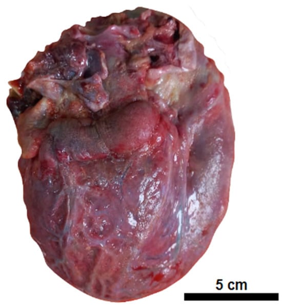
Myocarditis following COVID-19 mRNA vaccination: https://www.mdpi.com/2076-393X/12/2/194 Case of a 7-year-old boy. He died. "The heart exhibited a granular, opaque, whitish external surface, akin to fibrin deposits on the pericardium"

Fatal Myocarditis following COVID-19 mRNA Immunization: A Case Report and Differential Diagnosis Review Carditis in childhood is a rare disease with several etiologies. We report a case of infant death due to pericarditis and myocarditis after the mRNA vaccine against COVID-19 (COVIDmRNAV). A 7-year-old male child received the first dose of the COVIDmRNAV and presented with monoarthritis and a fever non-responsive to oral antibiotics. The laboratory investigation showed signs of infection (leukocytosis, high levels of c-reactive protein). His condition rapidly deteriorated, and the patient died. The autopsy identified pericardial fibrin deposits, hemorrhagic areas in the myocardium, and normal valves. A diffuse intermyocardial inflammatory infiltrate composed of T CD8+ lymphocytes and histiocytes was identified. An antistreptolysin O (ASO) dosage showed high titers. The presence of arthritis, elevated ASO, and carditis fulfills the criteria for rheumatic fever. However, valve disease and Aschoff’s nodules, present in 90% of rheumatic carditis cases, were absent in this case. The temporal correlation with mRNA vaccination prompted its inclusion as one of the etiologies. In cases of myocardial damage related to COVID-19mRNAV, it appears to be related to the expression of exosomes and lipid nanoparticles, leading to a cytokine storm. The potential effects of the COVID-19mRNAV must be considered in the pathogenesis of this disease, whether as an etiology or a contributing factor to a previously initiated myocardial injury. mdpi.com
Saved - February 27, 2024 at 2:53 PM

@DrJohnB2 - Dr John B.

(2/n) Example 2 https://t.co/pivGY1yVSr

View Full Interactive Feed