reSee.it - Tweets Saved By @_HeartofGrace_

@_HeartofGrace_ - Christie Laura Grace
My name is Christie Grace. I'm a former biotech manager and coordinator, custom development, working with RNA and LNP to produce recombinant proteins and more. I left the industry for a few reasons. Unrelated to this, in 2021, I discovered people were being subjected to an experiment. I couldn't believe what was occurring. They were being experimented on. I was horrified. I reported the activities and the experiment where this was occurring, and I was ignored, except by a couple of people, but they weren't in positions of authority to do anything. They reported with me. Nothing was done. After a month or so, something happened which I can only speculate as to the reasons why, were they paid off? I don't know. But they went silent. So, I started recording. At first I recorded audio only. I wasn't aware I could access wide angle camera lens recording devices or know I could buy them. That wouldn't matter soon, because I would be sent that equipment. I spoke with more than one lawyer who said it was legal to do so, and I was not breaking any laws. Upset that no action was being taken, and that the experiment was continuing, it was advised I reach out to some news stations. My name and contact information was spread to all that you all know of, including the whistleblower one that you see all of the hidden camera recordings posted of. This was 2021. If I have a contract with one of them, the details of the contract cannot be spoken of, due to the nature and contents of the contract itself. I was sent high end undercover recording equipment, multiple types, that were sent fed ex overnight. I was then sent links on how to operate the equipment and other links to shuttle the videos I was recording with audio back to that organization, alongside other documents. Things got worse where I was getting data, for me, when I reported to the federal government. This forced the organization in question to start an internal investigation, but they botched it on many levels, and I video recorded their law breaking that occurred when they even violated the rules of the investigation itself. I recorded for ten months. I was also given the software to record in other ways, like phone calls, zoom meetings, etc. I was then forced out of that area, and it would take a long time for me to find a lawyer who was even willing to take on such a complex federal case. I was told by almost every lawyer I spoke to that it was incredible the data I got out, but that I had and I quote, "touched the third rail." I even have the experiment itself--the protocol. I have it all. It was the beginning of 2022 when I joined Twitter and I was nervous to do so. Someone in these spaces that has quite a following of half a million followers, who had about twenty thousand just two years ago when I joined, was getting death threats and even had his arm broken (now you have to know who that is), who told me to use my real name here, and to keep going, and keep speaking out. I had multiple journalists following me several who still do to this day who have been waiting for me to produce this data. In December of 2021 my case would be accepted by the federal government after it made it through the second round of their legal team. A lawyer would accept my case simultaneously to represent me. The case with the federal government isn't in a civil court where money is being asked for. My case is with the federal government itself. None of the news agencies who I spoke to wanted to take the case because and here's the kicker, the majority said that they won't "shame their own". I recorded them saying that. I have lawyers in emails stating they won't shame their own and that they wouldn't represent me. December 2021 is when the case was accepted. Of course lockdowns were still happening to some extent and many organizations lost employees. I joined Twitter to release those files when the government makes its decision. It will set legal precedent in another area that several of you are tracking regardless if I win or lose. When that case started it was advised initially that I wipe myself from the internet. So I deleted almost all traces of me. Google. People search. I deleted almost everything I could. All my other social media. LinkedIn. I had so many contacts on LinkedIn from biotech it wasn't even funny. Gone in a blink. Facebook. Anything I had written. I went farther into search parameters, and placed requests to have my data and name removed. I became effectively, a ghost. But then I knew I had to come back to release this data. I knew I needed the internet to do so. So when I joined Twitter and I saw people talking improperly about RNA and LNP, I thought oh my gosh here we go, am I going to step into another arena? Is this really happening? This just can't be happening. And then I couldn't believe it. Mice. What the heck is a mouse army? I remember laughing to myself thinking this is bonkers. I initially came here to release data. I can't release data until the federal government makes its decision. No one is stopping me from releasing the files I have. However, I need to give the organization in question who committed what I feel are crimes, the chance to respond to the government with their word against my physical documentation and proof. I need to show what actually happened, but I have to wait for them to respond. This is typical in court cases. You don't show your hand. I have been accused by multiple members of the mouse army, doctors, scientists, podcasters, professors, and others in these spaces of being CIA, NSA, or the like. That is the most ridiculous thing I've ever heard. I've also been called a baby killer by the same people which is recorded and archived. I've been called the most terrible things over the course of the last year. I was told if I didn't have receipts what I was claiming had occurred did not. Well guess what. And some of them found an old case that is absolutely meaningless trying to point at it as if it is some kind of gotcha moment. I'll give you the gotcha moment right here. I'm a federal whistleblower. I'm not Edward Snowden level but I am a federal whistleblower. And we are going on 3 years now and this case is about to be decided. And it's going to set legal precedent in a huge arena that is one of the primary things being discussed in this current presidential election cycle. I have also speculated that might be why they are dragging their feet on the decision, because this will affect current civil rights cases. The case that I have has multiple components to it and it will impact the law going forward in this country. So when I started getting harassed and abused on this site and defamed by people who said they were my friend, do you know what I did? I gathered evidence. And I've been doing so since at least the last year. I'm horrified and disappointed at the level of abuse that I and others have received here from doctors, scientists, and others who said they were here to protect people. They just covered things up, just like what happened in 2021. Obfuscation These people are no different. There's no demarcation line. I have been fighting for 3 years simultaneously for the vaccine injured, against experimentation on humans, and for multiple levels of civil rights and human rights. And I have been sh1t on by people who lied who are doctors, scientists, and professors, who claim to have moral high ground. And I have had enough.

@_HeartofGrace_ - Christie Laura Grace
Because I've been threatened and harassed by people who seem to not care what they say publicly, about me, know that all of the files I have, the recordings, and the evidence and communications I have, have been physically sent to two people who are pretty known in these circles who I can trust. If anything happens to me, even if I get sick from this blood clot and get worse from this blood clot and land in ICU, all files will be released. Not just the files on the experimentation. The abuse and harassment of me.
@_HeartofGrace_ - Christie Laura Grace
1/ 💉LNP: Lipids and Liposomes: GENE DELIVERY to the PLACENTA: A STUDY. The placenta, like the heart, runs primarily on lipid metabolism. A thread on lipids used in "gene delivery", LNP entering the placenta, and what occurs when you disrupt lipid balance in the placenta.

@_HeartofGrace_ - Christie Laura Grace
2/ First study: "Liposomes as Gene Delivery Vectors for Human Placental Cells" https://www.ncbi.nlm.nih.gov/pmc/articles/PMC6099662/ doi: 10.3390/molecules23051085.


@_HeartofGrace_ - Christie Laura Grace
3/ The study employed an in vitro model using human primary villous cytotrophoblasts to evaluate the efficacy of liposomes in delivering siRNAs to placental cells. lipofectamine reagent, a commonly used transfection agent, was compared against cationic lipids (positive charge)
@_HeartofGrace_ - Christie Laura Grace
4/ "The outer cell layer, the syncytiotrophoblast (ST), is in direct contact with the mother’s blood flow containing the administered drugs." "microscopy observations showed that all tested formulations led to fluorescent siRNA uptake in the primary cytotrophoblasts."
@_HeartofGrace_ - Christie Laura Grace
5/ "Cytotrophoblasts continually differentiate into syncytiotrophoblasts during villous formation and development. Cytotrophoblast invasion into the uterine spiral arteries is accompanied by loss of the endothelial lining and musculoelastic tissue in these vessels."

@_HeartofGrace_ - Christie Laura Grace
6/ ( https://www.ncbi.nlm.nih.gov/books/NBK53245/) "Further experiments to study the toxicity of lipoplexes on VCT should be performed."

@_HeartofGrace_ - Christie Laura Grace
7/ Next: "Plastic and Placenta: Identification of Polyethylene Glycol (PEG) Compounds in the Human Placenta by HPLC-MS/MS System" DATE: online 2022 Oct 22. doi: 10.3390/ijms232112743 https://www.ncbi.nlm.nih.gov/pmc/articles/PMC9656682/
@_HeartofGrace_ - Christie Laura Grace
8/ "Although studies indicate the low toxicity of polyethylene glycol on living organisms [8], there are reports of nephrotoxicity [9] damage to the central nervous system and heart, as well as pulmonary and renal failure [10] in PEG-treated animals."
@_HeartofGrace_ - Christie Laura Grace
9/ 🚨 In the present study, we find evidence of PEG exposure in the human placenta, through mass spectrometry analysis. Liquid chromatography–MS/MS technology is a powerful and sensitive technique for the simultaneous characterization and separation of each PEG component [11].
@_HeartofGrace_ - Christie Laura Grace
10/🚨🚨 "We found the presence of PEG in ten out of twelve examined human placentas," "a series of polyethylene glycols (PEGs) and to demonstrate that this xenobiotic particle crosses the placental barrier."

@_HeartofGrace_ - Christie Laura Grace
12/ (Vicki Loves Facs?) Look closely--we are looking at the Chorioamniotic

@_HeartofGrace_ - Christie Laura Grace
13/ "In this study, high-resolution mass spectrometry was used to identify mono- and polymers of PEGs, therefore it is necessary to continue with a metabolomics analysis to investigate the influence on the metabolism by the low molecular weight polymers detected, comparing the
@_HeartofGrace_ - Christie Laura Grace
14/ "samples of the placenta in which the presence of high and low molecular weight PEG was detected with samples in which no plastic particles were detected.""
@_HeartofGrace_ - Christie Laura Grace
15/ "Histopathological studies of the placenta in IUGR indicate that abnormalities of the maternal spiral arterioles, dysregulated villous vasculogenesis, and abundant fibrin deposition are characteristic in IUGR (Redline, 2008)."
@_HeartofGrace_ - Christie Laura Grace
16/ "Intrauterine growth restriction, human placental development and trophoblast cell death" https://www.ncbi.nlm.nih.gov/pmc/articles/PMC2742274/#:~:text=a%20normal%20pregnancy.-,Histopathological%20studies%20of%20the%20placenta%20in%20IUGR%20indicate%20that%20abnormalities,IUGR%20(Redline%2C%202008).

@_HeartofGrace_ - Christie Laura Grace
17/ "In summary, our study shows a profound alteration in the placenta of IUGR patients with respect to energy and lipid metabolism," . doi: 10.3390/biomedicines10061411. https://www.ncbi.nlm.nih.gov/pmc/articles/PMC9220006/
@_HeartofGrace_ - Christie Laura Grace
18/ LNPs contains lipids (liposomes) Depending on the amount entering, exogenous liposomes could interfere with the normal exchange of nutrients and oxygen between the mother and the fetus, negatively impact fetal development and growth.
@_HeartofGrace_ - Christie Laura Grace
19/ 🚨💉 "vesicle-like nanocarriers such as liposomes and exosomes can also enter the placental barrier through membrane fusion." https://www.tandfonline.com/doi/full/10.1080/10717544.2023.2184315

@_HeartofGrace_ - Christie Laura Grace
20/ "significant translocation of liposomes inside the placenta." https://hal.science/hal-03712174/document
@_HeartofGrace_ - Christie Laura Grace
21/ "In-Utero Neurotoxicity of Nanoparticles" https://www.intechopen.com/chapters/79886
@_HeartofGrace_ - Christie Laura Grace
22/ "However, other polymeric NPs for gene delivery that use highly cationic molecules such as polyethyleneimine (PEI) have been found to be highly toxic" "Additionally, LNPs contain an ionizable lipid component that remains uncharged at neutral pH
@_HeartofGrace_ - Christie Laura Grace
23/ (they left out the charged ones in the article. There are charged particles and the LNPs are not neutral. )
@_HeartofGrace_ - Christie Laura Grace
24/ "Immune activation by nucleic acids: A role in pregnancy complications" Cell-free self-DNA or RNA may induce an immune response by activating specific sensing receptors. During pregnancy, placental nucleic acids present in the maternal circulation
@_HeartofGrace_ - Christie Laura Grace
25/ further activate these receptors due to the presence of unmethylated CpG islands. A higher concentration of cell-free foetal DNA is associated with pregnancy complications and a higher risk for foetal rejection https://onlinelibrary.wiley.com/doi/10.1111/sji.12651
@_HeartofGrace_ - Christie Laura Grace
🚨 🚨🚨MODERNA (CATALENT MAKES MODERNA) "a total of 179 complaints were received for particles (foreign matter, particulate matter, black specks, black particles, foreign particle, black impurities. black dots and other particulates, dark particles, foreign material, foreign object, dark gray particulate, or pieces" "Your firm does not document the identity of particulates found during visual inspection of aseptically filled product." 'YOUR FIRM FAILED TO THOROUGHLY INVESTIGATE ANY UNEXPLAINED DISCREPANCY OR FAILURE OF A BATCH OR ANY OF ITS COMPONENTS TO MEET ANY OF ITS SPECIFICATIONS, WHETHER OR NOT THE BATCH HAS ALREADY BEEN DISTRIBUTED" !!!!!! --BATCHES WERE RELEASED ANYWAYS AFTER EXCEEDING VISIBLE PARTICLES TESTING --particles identified not even used in manufacturing processes --cleaning has yet to be updated --Batches that exceeded internal control limits not always investigated and released anyways --foreign particles (polypropylene and rubber) --replacement and interventions not performed in timely manner --Multiple manufacturing events/deviations --a total of 52 supplier complaints for stopper related issues --Your firm did not adequately extend your complaint investigation to other batches of the same lot or product when warranted. --high number of complaints reported for the same defect for the same product manufactured at the firm --Control procedures are not established which monitor the output and validate the performance of those manufacturing processes that may be responsible for causing variability in the characteristics of inprocess material and the drug product. --Acceptance criteria for the sampling and testing conducted by the quality control unit is not adequate to assure that batches of drug products meet appropriate statistical quality control criteria as a condition for their approval and release. --Procedures designed to prevent microbiological contamination of drug products purporting to be sterile are not established, written, or followed. !!!!-Adequate justification could not be provided for not performing contact plating o --The emulsion visual inspection kits do not represent extrinsic particulate matter. --You cannot confirm through documentation the inspection times during the execution of visual inspection qualification tests is representative of routine visual inspections --There is no scientific justification for not performing particles testing on routinely inspected product --Procedures designed to prevent microbiological contamination of drug products purporting to be sterile did not include adequate validation of the aseptic process. --Records are not maintained so that data therein can be reviewed at least[o)-(4) J to evaluate the quality standards of each drug product to determine the need for changes in specifications or manufacturing or control procedures. --Your firm did not establish criteria during supplier qualification and requalification based on recurring supplier complaints in A-SOP-03-03-003, https://www.fda.gov/media/162418/download
@_HeartofGrace_ - Christie Laura Grace
1/ 🧵💉Adverse Events: All of the threads combined on RNA/DNA/LNP. People have been asking for easier to comprehend posts. I am not one to drink, but consider this thread "science in a shot glass" All of the threads have study links to back what is stated, and explanations.

@_HeartofGrace_ - Christie Laura Grace
2/ The LNP has a charge. It can have a neutral charge, positive charge (+) or negative charge (-). The RNA and DNA have a negative charge (-). The ionizable lipids have a positive charge (+). Charge determines where in the body it goes and what it does.

@_HeartofGrace_ - Christie Laura Grace
9/ If the charge is positive (+), it is zipping right to the lungs. It does not matter if you aspirate. This is by injecting into the MUSCLE, not just IV. MUSCLE. It could cause injury in lung (PE?). If a little negative, it goes to spleen. If VERY negative, it goes to HEART.

@_HeartofGrace_ - Christie Laura Grace
3/ There is net charge (positive and negative charges added up--similar to if you add 3 + -5 = -2 ). That is it's net charge. Zeta potential is the kinetic charge (a potential of interaction) Positively charged LNPs cause MASSIVE clots in the lungs.

@_HeartofGrace_ - Christie Laura Grace
4/Negatively charged LNPs can enter the cardiomyocyte (10:1 ratio (+) LNP to (-) RNA/DNA) and cause MYOCARDITIS. The DNA contamination changed the charge on the LNP. The DNA is very negatively charged. The scientists involved should have known this.

@_HeartofGrace_ - Christie Laura Grace
21/ DNA in the LNP, and changed the ration form 20:1 lipids to DNA plasmid, to 10:1 DNA to lipids, the cardiomyocyte was transfected. As a reminder, they were not using a bulk plasmid with an SV40 promoter on it, that can express the spike protein, and other contaminants.

@_HeartofGrace_ - Christie Laura Grace
5/ Highly negatively charged LNP can cause clots. This will drive clot formation thousands of times more aggressively than human body normally would. It makes clots form more quickly, with more fibrin (thick and spindly), and are more difficult to treat.

@_HeartofGrace_ - Christie Laura Grace
🚨 Higher negative charges on the LNP can cause clots. "Influence of liposome charge and composition on their interaction with human blood serum proteins" The LNP contains positively charged lipids, negatively charged RNA, and recent DNA contamination which carries a negative charge. The presence of the DNA plasmid contamination, is throwing off the charge on the LNP, causing it to be more negative. The tests the companies did on the LNP when stated they had a neutral charge was not with the introduction of DNA plasmid contamination, it was a different process. This would not matter anyways, the charge and zeta potential shifts negative by 7 clicks right as it enters the human body when it hits the blood as compared to what it is in a bulk solvent. This does not account for interactions with proteins that have a negative charge, or other charged elements, driving it further negative. Negatively charged LNPs can cause damage, including injuring the lining of endothelium, and clots. https://link.springer.com/article/10.1007/BF00926084

@_HeartofGrace_ - Christie Laura Grace
6/ The LNP can cross the placenta, and "transfect" it--meaning it can enter the cells there, and growing baby. There is a 100 study 124 tweet thread I made on this. 100+ studies exist on it. This is not a hypothesis. The LNP crosses the placenta.

@_HeartofGrace_ - Christie Laura Grace
7/ The plasmid DNA contamination, can cause the body to attack itself, temporarily or indefinitely. The body sees it as foreign, but also, it could see it as "self", causing the body to attack the tissues the LNP enters and create auto immune situation.

@_HeartofGrace_ - Christie Laura Grace
1/🧵:Re: DNA Plasmid Contamination: (only focusing on this--yes all other risks): ONE of MANY IMMUNE responses may be due to one part of plasmid--body recognizes as foreign (bacteria/viral similarity), and launches an attack: the unmethylated CpG motifs in DNA in the plasmid

@_HeartofGrace_ - Christie Laura Grace
8/ The DNA plasmid contamination may disrupt the normal growth of a baby, causing an auto immune response in the baby, issues with pregnancy, delivery, defects, and other concerns.

@_HeartofGrace_ - Christie Laura Grace
1/ 🚨🧵Gestational exposure to unmethylated CpG ODN (found in DNA plasmid Contamination in modRNA "vaccines") CAN DISRUPT growth and development of both the fetus (baby) and placenta. Additionally, it causes disruptions the daily rhythm of the mother's blood pressure." And😡:

@_HeartofGrace_ - Christie Laura Grace
9/ Sperm cells can spontaneously "take up" pieces of DNA. Plasmid DNA exists as contamination inside the LNP. If this enters the testes, and reaches the sperm, the sperm can absorb the DNA plasmid, altering it.

@_HeartofGrace_ - Christie Laura Grace
🚨💉🧬Mature sperm cells have the spontaneous ability to take up exogenous DNA. c@vid modRNA vaccines contain DNA plasmid (exogenous) -NO LNP or transfection agent needed--right into the nuclei (multiple studies. Here is one--no thread tonight) https://pubmed.ncbi.nlm.nih.gov/11139334/


@_HeartofGrace_ - Christie Laura Grace
10/ DNA plasmid contamination in modRNA "vaccines" DO NOT HAVE TO ENTER THE NUCLEUS to impact GENE EXPRESSION, and be implicated in CANCER RISK! The DNA plasmid interacts with different areas i the body, and can impact what genes do.

@_HeartofGrace_ - Christie Laura Grace
11/ The freezing and thawing process is braking the LNPs. it is causing some to leak out. It is also causing some to stick together, expand, and cluster, which can cause blockages. It can cause the RNA inside to break, which can be oncogenic.


@_HeartofGrace_ - Christie Laura Grace
20/ Another thing that could be happening, in addition to DNA plasmids changing the zeta potential (charge) is the LNP is breaking down during the freeze/thaw process, and parts can leak out, changing the overall charge as well. Dr. Ko and De, and others proved.


@_HeartofGrace_ - Christie Laura Grace
12/ Impurities in Positively Charged Lipids in the LNP, can MUTATE mRNA in LNP (Packer et al., 2021); potentially mutating other nucleic acids (RNA,DNA) that may cause: -mutation -Aberrant Protein (toxic) -Noncoding (can be oncogenic) -Misfold/aggregation

@_HeartofGrace_ - Christie Laura Grace
1/ Impurities in Positively Charged Lipids in the LNP, can MUTATE mRNA in LNP (Packer et al., 2021); potentially mutating other nucleic acids (RNA,DNA) that could lead to: -Point mutation -Aberrant Protein (toxic) -Noncoding (can be oncogenic) -Misfold that protein/aggregation

@_HeartofGrace_ - Christie Laura Grace
13/ When the LNP is made, with the RNA inside of it, not all of it is whole. Some of the RNA is in pieces when it is first made. Some of the whole pieces also break when it is frozen. Pieces of RNA can cause cancer or turn cancer off.

@_HeartofGrace_ - Christie Laura Grace
6/ This can cause other structures to form, aggregation, misfolding of proteins, non-coding of the mRNA (oncogenic) and mutations in nucleic acids they would interact with (RNA/DNA) and cause unwanted effects. https://www.the-scientist.com/features/long-noncoding-rnas-and-microproteins-can-spark-cancer-or-sometimes-squelch-it-70961
@_HeartofGrace_ - Christie Laura Grace
14/ "Over 100 different ionizable lipid chemistries were examined, all of which produced measurable levels of mRNA-lipid adducts. This indicates that the formation of these adducts is not limited to a specific type of ionizable lipid but is a broad class effect." ALL MUTATE.
@_HeartofGrace_ - Christie Laura Grace
15/ Positively charged lipids DAMAGE BACTERIA. Bacteria exists in humans in a ratio of 2:1 --bacteria to human cells. Lipid Nanoparticles used in modRNA "vaccines", contain positively charged lipids. The LNPs are going everywhere, including the gut https://t.co/sFjeZgKDKT https://t.co/dmAIi0q1z3

@_HeartofGrace_ - Christie Laura Grace
3/ Zeta potential of E. coli and S. aureus were found to be −44.2 and −35.6 mV, respectively. An additional layer of negatively charged LPS in Gram-negative bacteria, has been attributed to the higher negative potential of E. coli than that of S. aureus. Gram positive bacteria https://t.co/friK40VIMx

@_HeartofGrace_ - Christie Laura Grace
16/ Liver Cancer: dsDNA is implicated in Hepatocellular Carcinoma. dsDNA is part of DNA plasmid contamination discovered in💉. dsDNA is activated by the STING pathway, causing an immune system cascade, which can cause inflammation, injury, and cancer. https://t.co/8RCNpawgnz https://t.co/mWNc5TSQKy

@_HeartofGrace_ - Christie Laura Grace
1/ 🚨🧵🧬💉Liver Cancer: dsDNA is implicated in Hepatocellular Carcinoma dsDNA is part of DNA plasmid contamination recently discovered in the 🧬💉 dsDNA is activated by the STING pathway, causing an immune system cascade, which can cause inflammation, injury, and cancer. https://t.co/Nvlsp3VL3e

@_HeartofGrace_ - Christie Laura Grace
17/ Thyroid damage and auto immune disorders are linked to dsDNA. dsDNA is found in the current plasmid DNA contamination. This is slightly different in the thyroid ( histone H2B) Study + cases on 💉injury. https://t.co/stvu10n6rP https://t.co/OTbWe9R387

@_HeartofGrace_ - Christie Laura Grace
1/ 🚨🧵🧬💉 Second verse, same as the first. Thyroid damage and auto immune is linked to dsDNA. dsDNA is found in the current plasmid DNA contamination. This is slightly different in the thyroid ( histone H2B) Study + cases on 💉injury. https://t.co/EF73pSRqVi

@_HeartofGrace_ - Christie Laura Grace
18/ MYOCARDITIS (DNA plasmid contamination was recently found in c@vid modRNA "vaccines") in ANIMAL STUDIES CAUSES: MYOCARDIAL INFLAMMATION IN MURINE HEARTS after exposure and damages the CARDIOMYOCYTE https://t.co/BlniROZkuW https://t.co/Lsg0DBWNbo

@_HeartofGrace_ - Christie Laura Grace
How bad is my batch: Batch # FM7380 High DNA plasmid contamination correlated to VAERS--How Bad is my Batch dot com: High Myocarditis numbers. (look at the arrows) @drdrew @DrJBhattacharya https://t.co/edVV22XNWU


@_HeartofGrace_ - Christie Laura Grace
19/ How the DNA plasmid contamination is causing the LNP to bind with proteins and cause other harms in the human body. Speicher and friends found the higher the DNA plasmid contamination, the worse the SAE on record. https://t.co/eMNcQmvSjM https://t.co/OkFXWol2pW


@_HeartofGrace_ - Christie Laura Grace
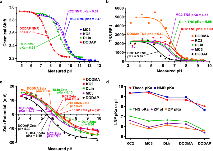
11/ Pf1zer states very clearly, in their own document, that they measured the LNP zeta potential to be closer to neutral, to "avoid non specific binding events in the blood compartment". THEY KNEW. They knew that if this would swing positive or negative, this would be bad. https://t.co/rwjMZU93xa

@_HeartofGrace_ - Christie Laura Grace
20/ Could the LNPs alter/impair the mononuclear phagocyte system (MPS), leading to toxicity, reduced pathogen clearance, impaired immune function, and tumor/cancer progression? Studies show a negative impact after repeated injections of lipids. https://t.co/lkVrKzAKsV https://t.co/yzjVSf6JdV

@_HeartofGrace_ - Christie Laura Grace
1/ 🧵Could Lipid Nanoparticles (LNPs) alter/impair the mononuclear phagocyte system (MPS), leading to toxicity, reduced pathogen clearance, impaired immune function, and tumor/cancer progression? First, an overview of the MPS, AKA, the reticuloendothelial system (RES): https://t.co/1sGrcn1oT0

@_HeartofGrace_ - Christie Laura Grace
21/ Spike protein is known to misfold and aggregate. When you make a recombinant protein in a lab, in cells, under very controlled conditions (this is when you introduce RNA inside and LNP in a cell even, no human--it can misfold). https://t.co/Z4I6SuiYP2

@_HeartofGrace_ - Christie Laura Grace
22/ Some LNP could not have formed properly in ways that may not have been explained, due to the DNA plasmid contamination. This just makes things far worse, and adds to the buffet of existing bad. LNPs weren't the only thing forming in the mix. https://t.co/UcmnmjVBca https://t.co/3DNRKhMMYK

@_HeartofGrace_ - Christie Laura Grace
23/ RAPID AAD-Aortic Aneurysm and Dissection (tearing/rupture) is lethal. Plasmid DNA is dsDNA. dsDNA can enter cells from the body's own sources, or a contamination event (💉) dsDNA is shown to cause AAD https://t.co/Wr1hCy9UIL https://t.co/EnC0nzL2gX

@_HeartofGrace_ - Christie Laura Grace
1/ 🚨🧵 RAPID AAD-Aortic Aneurysm and Dissection (tearing/rupture) is lethal. Plasmid DNA is dsDNA. dsDNA can enter cells from the body's own sources, or a contamination event (💉) dsDNA is shown to cause AAD via the STING path. A thread on a study, and adverse event cases. https://t.co/ylFT5y88jJ

@_HeartofGrace_ - Christie Laura Grace
24/ How Zeta Changes in the Human Body (layman's), and Why Pharma Companies are Incorrect Stating their Calculation of Zeta Potential (surface charge) is Accurate, much less, that it will not do harm. Zeta is the starting point for most adverse events. https://t.co/W0bs0jhNPZ https://t.co/zDBA0MlKPc

@_HeartofGrace_ - Christie Laura Grace
PURE ZETA: PART SEVEN: LNP: How Zeta Changes in the Human Body (layman's), and Why Pharma Companies are Incorrect Stating their Calculation of Zeta Potential (surface charge) is Accurate, much less, that it will not do harm. Zeta is the starting point for most adverse events. https://t.co/ni6d38C7PQ

@_HeartofGrace_ - Christie Laura Grace
1/ 🚨🧵 RAPID AAD-Aortic Aneurysm and Dissection (tearing/rupture) is lethal. Plasmid DNA is dsDNA. dsDNA can enter cells from the body's own sources, or a contamination event (💉) dsDNA is shown to cause AAD via the STING path. A thread on a study, and adverse event cases. https://t.co/ylFT5y88jJ

@_HeartofGrace_ - Christie Laura Grace
ZETA POTENTIAL DRIVES CLOTS (COAGGULATION) AT DIFFERENT RATES IN DIFFERENT VESSELS (RABBIT STUDY) Different vessels showed significant differences in blood coagulation. The coagulation process was fastest in the inferior vena cava and portal vein and slowest in the renal vein, abdominal aorta, and renal artery. These differences in blood clotting correlated with the values of the zeta-potential measured in the vessels (r = 0.902). Factors affecting zeta-potential include the velocity of fluid movement, dielectric constant of the solution and contact surface, pH of the solution, and other factors known to influence the total electrokinetic potential. These differences are attributed to the metabolic activity of the vessel wall, the concentration of vasoactive substances in the blood, nerve supply to the blood vessels, and the local concentration of pro- and anticoagulants in the vessel wall and blood. Local heparin concentration in the vessel wall and blood also plays a role in zeta-potential, with high heparin concentration correlating with slow coagulation. Aorta and renal vessels have a high zeta-potential and slow coagulation, likely due to the presence of high heparin concentration.

@_HeartofGrace_ - Christie Laura Grace
According to my calculations (which I am not posting until I get confirmation from someone, at least one coming close to the charge that I got)--if you inject someone with 43 million lipid nanoparticles based on the new zeta potential, NO MATTER WHAT IS INSIDE: DNA, RNA, or just lipids, the following can occur: (sound familiar?) Immunological Response:The immune system may respond to the presence of foreign lipid nanoparticles, potentially leading to an immune reaction, inflammation, or immune cell activation. Blood-Brain Barrier penetration Oxidative Stress Endothelial Dysfunction Thrombocytopenia Hematological Effects Renal Implication Respiratory Effects Disruption of Cellular Signaling Metabolic Disturbances Genotoxicity Reproductive and Developmental Impact Death Coma Stroke Heart Attack Seizure Autoimmune Clotting and Cardiovascular Effects Inflammatory Response Tissue Distribution, Accumulation, and DAMAGE Cellular Damage Cytotoxicity Organ Function Systemic Toxicity Hemodynamic Consequences This is all without any impact of RNA or DNA. This is just the zeta potential pushing far negative as a result OF the DNA. Not an interaction with it. That is separate. RNA is separate concern. Spike is separate concern. ALL of this would happen, based on the new zeta calculation I got based off 200 billion pieces of DNA plasmid entering 43 million LNP at a size of 100-12 base pairs.
@_HeartofGrace_ - Christie Laura Grace
1/ This video shows the reaction which occurs if you expose an amount of the Pf1zer vaccine to human blood. If this happened in real life, to a human, that person would drop over. Many components are missing here. Let's go over the reaction first. (zeta) https://rumble.com/v269jdu-pfizer-genetic-vaccine-added-directly-to-human-blood.html

@_HeartofGrace_ - Christie Laura Grace
2/ (This is with much respect to all involved, however, incomplete data is being shared. If I stand alone, so be it.) Human blood, in a health person, who does not smoke, who does not take blood thinners, has a pH and contains many charged particles. Red blood cells, are
@_HeartofGrace_ - Christie Laura Grace
3/ also called erythrocytes. Erythrocytes have a zeta potential and that changes, depending on where they are (they would have a different zeta if present in urine, for example--see the dozen threads I made on this). In human blood, red blood cells have an average zeta https://t.co/LcpOy1wQ1N

@_HeartofGrace_ - Christie Laura Grace
4/ of around −15.7 mV, but this can vary depending on person, location in the body, and in the surrounding environment. What was done in this video, is that the contents of a vaccine were placed on a slide with blood. For instance, different blood vessels have different
@_HeartofGrace_ - Christie Laura Grace
5/ sizes, which influence fluid dynamics, shear forces, and zeta potential within that area, which is different than the zeta potential on a particle. Different blood vessels also exhibit distinct hemocoagulation patterns. Vessels w/ slower blood coagulation have higher
@_HeartofGrace_ - Christie Laura Grace
6/ zeta-potential values. Differences in content of pro- and anticoagulants in the vessels contribute to variations in blood coagulation. Also, local heparin concentration in the vessel wall and blood also plays a role in zeta-potential, with high heparin concentration
@_HeartofGrace_ - Christie Laura Grace
7/ correlating with slow coagulation. The aorta and renal vessels have a high zeta-potential and slow coagulation, due to presence of high heparin concentration. The high viscosity of blood in the portal vein and its low zeta-potential indicate relative hypercoagulation, which
@_HeartofGrace_ - Christie Laura Grace
8/ corresponds to increased blood viscosity. As stated in the other threads on zeta, the zeta of a particle is different regarding its overall net charge when it is in blood, compared to CSF, compared to lung (lung has high negative charges due to negatively charged mucins
@_HeartofGrace_ - Christie Laura Grace
9/ in the mucous, as well as differences in pH, ions, viscosity, and molecules along with flow. Regarding this one slide, there is no blood vessel wall, shear force, immune system, flow, or other interactive forces that will come into play. This is just on a slide.
@_HeartofGrace_ - Christie Laura Grace
10/ and if this happened in rea life, inside your vein, you would be on a gurney. Now regarding what happened in this video. We know that pfizer measure the zeta potential while in bulk solvent of a lipid nanoparticle containing only RNA, positively charged lipids, dspc, peg,
@_HeartofGrace_ - Christie Laura Grace
11/ and cholesterol as being -3 millivolt in a bulk solvent that was free of ions, at a specific pH, in a thinner solution, which is much different than the environment in the human body, which has its own localized areas of zeta and electrical fields which all impact kinetics. https://t.co/jAz2Cp0rve

@_HeartofGrace_ - Christie Laura Grace
12/ If an LNP contains DNA plasmids, these things have a high negative charge as well, so depending on how many landed in each LNP, that means you are adding negative to negative, which would make the overall net charge of the LNP to be even MORE negative (for zeta explanation--
@_HeartofGrace_ - Christie Laura Grace
13/ read the 14 threads--this will not be covered here). We will assume based on calculations that I already did, that if you have 43 million LNP per shot, with about 120 average base pair, when calculating the phosphates, without the addition of more positively charged lipids
@_HeartofGrace_ - Christie Laura Grace
14/ because those are a fixed amount, with an average of 200 billion pieces of DNA plasmid per shot, and if the zeta potential was measured as -3 millivolt without any plasmids in a bulk solvent, then approximate guess (with algebra assist), if we take -3 millivolt, and then
@_HeartofGrace_ - Christie Laura Grace
15/ assume one shot has 43 million LNP and distribute 200 billion pieces of DNA at an average of 120 base pair, and recalculate zeta for the LNP IF IN ETHANOL, the new approx. zeta is IMPOSSIBLE. Everyone would have died taking this shot. Because if you evenly distribute, you
@_HeartofGrace_ - Christie Laura Grace
16/ have 4651.16 pieces per LNP. So let's just assume -40 millivolt (it is higher--it is crazy higher). New zeta is -40 (it's not--it's higher, this is conservative). Only on a slide with blood, the reason the video shows the blood (red blood cells have negative charge) are
@_HeartofGrace_ - Christie Laura Grace
17/ moving away from the vaccine, is because the LNP have a high negative charge, and red blood cells also have a high negative charge, so repulsion will occur, however, that is not the only thing occurring. We have negative zeta blood cells interacting with negative charged LNP
@_HeartofGrace_ - Christie Laura Grace
18/ You have aggregation. Reasons are: Differences in local charge density: Charge distribution on blood cells are not always uniform, thus some areas of the blood have a higher or lower charge density, leading to variations in the electrostatic interactions with the negatively
@_HeartofGrace_ - Christie Laura Grace
19/ charged LNPs. In areas where charge density is lower or where repulsive forces are weaker, there is a higher chance of aggregation. This could occur if the LNPs can overcome repulsion and come into close proximity to blood cells. Also, Blood cells have surface charge
@_HeartofGrace_ - Christie Laura Grace
20/heterogeneity--different cell types or different regions of individual cells have varying charge distributions. This heterogeneity changes the interactions with LNPs. Also, protein coronas can form FAST. Plasma proteins in the blood may adsorb onto the surface of the LNPs,
@_HeartofGrace_ - Christie Laura Grace
21/ altering their charge and behavior. The interactions between plasma proteins, nanoparticles, and blood cells contribute to aggregation and repulsion forces. Furthermore, ion bridging can occur, which I have spoken about in great detail. For example, divalent cations like
@_HeartofGrace_ - Christie Laura Grace
22/calcium (Ca2+) can contribute to ion bridging and facilitate aggregation by interacting with negatively charged surfaces, like the LNP and red blood cells, creating a bridge between them (- + -) Also, near the isoelectric point of a particle or blood cell, the net charge is
@_HeartofGrace_ - Christie Laura Grace
23/ reduced, and electrostatic repulsion is weaker, potentially leading to aggregation. This was a concentrated amount of the vaccine that was placed on a slide with blood. Shear force is not present, changes in viscosity, pressure, other ions, zeta potential of local areas,
@_HeartofGrace_ - Christie Laura Grace
24, movement of molecules, permittivity of free space, dielectric constant of the medium (blood or surrounding environment), dynamic viscosity, changes in pH, temperature, other ions present, ions in vascular walls proteins in vessels and organs, drugs a person takes, etc.
@_HeartofGrace_ - Christie Laura Grace
@_HeartofGrace_ - Christie Laura Grace
1/ 🚨👀🚨🧵Animal models show exposure to CpG ODN (this is also present in the DNA PLASMID contamination in the modRNA "vaccines") can cause autoimmune disease. A Case Study: rats and AUTOIMMUNE UVEITIS.

@_HeartofGrace_ - Christie Laura Grace
1/🧵:Re: DNA Plasmid Contamination: (only focusing on this--yes all other risks): ONE of MANY IMMUNE responses may be due to one part of plasmid--body recognizes as foreign (bacteria/viral similarity), and launches an attack: the unmethylated CpG motifs in DNA in the plasmid

@_HeartofGrace_ - Christie Laura Grace
2/ TWO different T cell ligands can cause synergistic activation of T cells. CpG-ODNs are known to be strong inducers of innate immunity and promote the generation of adaptive immunity. While autologous rat IRBP alone is not a strong stimulator for uveitogenic T cells,
@_HeartofGrace_ - Christie Laura Grace
3/ a robust T cell response is initiated when both autologous IRBP and CpG-ODN1826 are present. This suggests that the synchronized effect of these two molecules readily results in autoimmune uveitis.
@_HeartofGrace_ - Christie Laura Grace
4/ Autologous rat IRBP (interphotoreceptor retinoid-binding protein), "becomes" a potent uveitogen in the presence of small amounts of CpG-ODN1826. This combination leads to increased activation of uveitogenic T cells. CpG-ODN is needed to activate these T cells in vivo.
@_HeartofGrace_ - Christie Laura Grace
5/ CpG-ODN the activates autoreactive T cells both in vitro and in vivo. This alteration in the immune response impacts the pathogenesis of autoimmune uveitis.
@_HeartofGrace_ - Christie Laura Grace
6/ Layman's terms: CpG-ODN plays a significant role in turning on and increasing the number of immune cells (T cells) that mistakenly attack the body's own tissues. This happens both in laboratory settings (in vitro) and in living organisms (in vivo).
@_HeartofGrace_ - Christie Laura Grace
7/ The highly activated immune response is linked to the development of autoimmune uveitis, a disease where the immune system attacks the eye, causing inflammation and vision problems. CpG-ODN is the driver behind this autoimmune eye disease.
@_HeartofGrace_ - Christie Laura Grace
8/ The exact mechanisms by which CpG-ODNs enhance T cell activation in vivo and in vitro seems to be due to activation of antigen-presenting cells (APCs). CpG-ODN1826 appears to decrease the threshold for T cell activation, by facilitating interactions between different immune
@_HeartofGrace_ - Christie Laura Grace
9/ cells, leading to increased activation of autoreactive T cells. CpG-ODN lowers the level of activation needed for T cells to "get going". CpG is helping different types of immune cells "talk to each other", which results in the immune system getting more excited and mistakenly
@_HeartofGrace_ - Christie Laura Grace
10/ attacking the body's own tissues, as in autoimmune diseases. THIS is what we call molecular mimicry. cells) that mistakenly attack the body's own tissues. CpG-ODN1826 drives how this autoimmune eye disease happens.
@_HeartofGrace_ - Christie Laura Grace
11/ CpG-Containing Oligodeoxynucleotide 1826 Converts the Weak Uveitogenic Rat Interphotoreceptor Retinoid-Binding Protein Peptide 1181–1191 into a Strong Uveitogen 1 Shao et el J Immunol (2003) 171 (9): 4780–4785. https://doi.org/10.4049/jimmunol.171.9.4780
@_HeartofGrace_ - Christie Laura Grace
@_HeartofGrace_ - Christie Laura Grace
13/ TL;DR: the study focused on rats' eyes to investigate autoimmune uveitis, an inflammatory eye disease, to understand how the presence of bacterial DNA, specifically CpG-ODN1826, influences the activation and expansion of immune cells, particularly autoreactive T cells
@_HeartofGrace_ - Christie Laura Grace
14/ , in the context of autoimmune uveitis. The study found that CpG-ODN1826 substantially drives the immune response, leading to autoimmune uveitis, a condition where the immune system mistakenly attacks the eye's tissues.
@_HeartofGrace_ - Christie Laura Grace
15/ https://www.ncbi.nlm.nih.gov/books/NBK459445/

@_HeartofGrace_ - Christie Laura Grace
16/ "The bovine and rat IRBP peptides (Fig. 1 A) were synthesized by ResGen Invitrogen (Carlsbad, CA). CpG-ODN1826 (5′-TCCATGACGTTCCTGACGTT-3′), and the control ODN1982, a non-CpG-containing ODN (5′-TCCAGGAGTTCTCTCAGGTT-3′) (25) were purchased from Qiagen (Alameda, CA); the LPS
@_HeartofGrace_ - Christie Laura Grace
17/ "content of the ODN was <1 ng LPS per mg of DNA, measured using the Limulus amebocyte assay (BioWhittaker, Walkersville, MD)."
@_HeartofGrace_ - Christie Laura Grace
@DrJBhattacharya @drdrew (I hope you do not mind I am patterning you two gentlemen in my tweets) @drcole12
@_HeartofGrace_ - Christie Laura Grace
"partnering" Ugh! Silly phone. Happy Friday
@_HeartofGrace_ - Christie Laura Grace
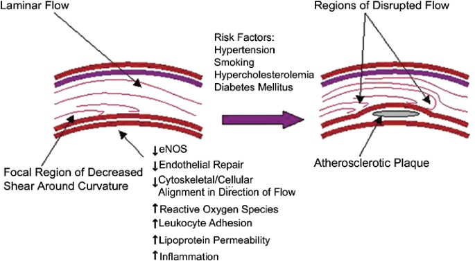
1/ 🚨🧵BLOOD VESSELS, Spike Protein, LNP, Immune System and CLOTS: PART TWO: THE LINING OF BLOOD VESSELS CARRY A CHARGE, AND CAN CHANGE WHEN INFLAMMATION AND INJURY OCCURS. Many interesting studies on animals occurred in the 80s, and this is another. (zeta on LNP will matter)

@_HeartofGrace_ - Christie Laura Grace
1/ 🚨🧵BLOOD VESSELS, Spike Protein, LNP, Immune System and CLOTS: PART ONE: Blood Vessels 101. Several studies on charges (+/-) of vessels, blood, and the harm lipids can do were performed in the 1980s. They are still relevant. Unearthed a very important one today.

@_HeartofGrace_ - Christie Laura Grace
2/ The study: Electronic Antihemocoagulation DeLangis, P. A., & Yen, T. F. (1986). Electronic antihemocoagulation. Biomaterials, medical devices, and artificial organs, 14(3-4), 195–225. https://doi.org/10.3109/10731198609117543 https://pubmed.ncbi.nlm.nih.gov/3814714/
@_HeartofGrace_ - Christie Laura Grace
3/ The study asks whether electrical current through blood and vessels can either extend the time it takes for blood to clot or prevent clot formation. The study aims to identify at what point in the clotting process these effects occur. This was done with and without animals.

@_HeartofGrace_ - Christie Laura Grace
4/ All cells and surfaces in the body carry an electrical charge, influenced by characteristics of cells, particles, and surrounding medium (liquid or solid). In the vascular system (blood vessels, heart, etc), most particles in the blood carry a NEGATIVE CHARGE.

@_HeartofGrace_ - Christie Laura Grace
5/ Red blood cells are negatively charged due to the presence of negatively charged sialic acid residues on their surface glycoproteins. The zeta potential of red blood cells falls in the millivolt (mV) range, with values ranging from -10 mV to -30 mV under physiological
@_HeartofGrace_ - Christie Laura Grace
6/ conditions. These values can change depending on factors like pH and the presence of other ions in the blood. White blood cells also carry a negative charge, primarily due to the negatively charged sialic acid residues on their surface glycoproteins. https://www.semanticscholar.org/paper/Electrical-properties-of-the-red-blood-cell-and-Fernandes-C%C3%A9sar/2f1a754b0cd2773c00bac9665db597290ee1fc66

@_HeartofGrace_ - Christie Laura Grace
7/ The zeta potential of white blood cells can vary between different types of leukocytes and under different conditions. The zeta potential of white blood cells can be affected by factors such as pH, ionic strength, and the presence of other ions in the blood. Additionally,
@_HeartofGrace_ - Christie Laura Grace
8/ the activation state of white blood cells (e.g., activated vs. resting) and their specific type can lead to variations in zeta potential. Platelets, like other blood cells, also have a negative charge on the surface, primarily due to the presence of negatively charged groups.

@_HeartofGrace_ - Christie Laura Grace
9/ : Blood plasma contains proteins, like albumin and globulins, which have both positive and negative charges. Albumin has a net negative charge, while some globulins may have a net positive charge. These proteins contribute to the overall zeta potential of the blood.

@_HeartofGrace_ - Christie Laura Grace
10/ The intima (inner layer) of blood vessels is typically negatively charged compared to adventitia (outer layer). However, trauma to the blood vessel can neutralize or even make the charge positive, leading to thrombosis (clot formation) at the injury site. The charge changes!

@_HeartofGrace_ - Christie Laura Grace
11/ If a cut is made into a blood vessel, it results in a POSTIVE CHARGE at the injury site. The study shows if the cut is kept negatively charged by applying an electrical current, clotting at the site will be inhibited, and the wound will continue to ooze. Conversely, if the
@_HeartofGrace_ - Christie Laura Grace
12/ electrical current is reversed and made positive, clotting will accelerate. When oppositely charged electrodes were submerged in a beaker of blood, a clot formed only at the positive electrode. Additionally, under similar conditions, white blood cells (leukocytes)
@_HeartofGrace_ - Christie Laura Grace
13/ migrated toward the negative electrode, indicating a change in cell polarity from negative to positive, possibly as a response to combat inflammation. https://www.researchgate.net/figure/The-leukocyte-recruitment-cascade-possible-effects-of-MPO-In-noninflamed-tissue_fig5_229555575


@_HeartofGrace_ - Christie Laura Grace
14/ In the vascular system, the intima is negatively charged compared to the adventitia. Vessel trauma can lead to a change in charge (neutral or positive) and result in thrombosis. This means the charge is moving from negative to positive, when inflammation and injury occurs
@_HeartofGrace_ - Christie Laura Grace
15/ in the lining of human blood vessels. "Vessel trauma" is any form of injury or damage to the blood vessel, caused by physical injury, surgical procedures, or disease-related damage.
@_HeartofGrace_ - Christie Laura Grace
16/ When a blood vessel experiences trauma, it can lead to a change in electrical charge. This change can manifest in two ways: Neutral Charge: The negative charge in the intima may become neutral, meaning it loses its excess negative charge. b. Positive Charge:
@_HeartofGrace_ - Christie Laura Grace
17/ In some cases, trauma can cause the negative charge to reverse and become positive.
@_HeartofGrace_ - Christie Laura Grace
18/ Thrombosis is blood clot formation within a blood vessel. A change in the electrical charge of the blood vessel's inner lining, particularly when it becomes neutral or positive due to trauma, is associated with the initiation or acceleration of the thrombosis process.
@_HeartofGrace_ - Christie Laura Grace
19/ "Negatively charged phospholipids, most particularly phosphatidylserine, are required for binding of the substrates, fIX or fX, to the phospholipid surface." https://www.ncbi.nlm.nih.gov/pmc/articles/PMC4826570/

@_HeartofGrace_ - Christie Laura Grace
20/ This is all going to come together in the following threads discussing the first waves of covid infection, DNA plasmid contamination with a high negative charge contaminating the current RNA "vaccine", and what happened to some people with infection vs. RNA "vaccination".
@_HeartofGrace_ - Christie Laura Grace
21/ It will also show why the negatively charged LNP, especially those with a higher negative charge which contain even more DNA plasmids, contributed to not only endothelial damage, but myocarditis. There are lot numbers here in this study which show higher DNA plasmid
@_HeartofGrace_ - Christie Laura Grace
22/ contamination led to higher rates of adverse events. If you look at each one of these lot numbers listed here in this study, you will see myocarditis as a primary severe adverse event, alongside clotting.

@_HeartofGrace_ - Christie Laura Grace
23/ I am going to bring you closer to what this mechanism should be. https://www.ncbi.nlm.nih.gov/pmc/articles/PMC8088814/

@_HeartofGrace_ - Christie Laura Grace
@DrJBhattacharya @drdrew
@_HeartofGrace_ - Christie Laura Grace
1/ 🧵Nattokinase: I do not take this for the reasons I do not (no need, also, not enough literature for me). But others DO take it. I do not promote its use here. However, this study shows nattokinase has a protective mechanism when it comes to the injury of BLOOD VESSELS.

@_HeartofGrace_ - Christie Laura Grace
2/ The study: https://www.lib.okayama-u.ac.jp/www/acta/pdf/64_6_399.pdf The researchers ni the study investigate the potential protective effects of natto extract on vascular endothelial damage induced by laser irradiation. Both the earlier waves of Covid and vaccination have pointed to endothelial injury.

@_HeartofGrace_ - Christie Laura Grace
3/ This study (separate on covid and endothelial injury) says : Alveolar-capillary endothelial cells can be activated by SARS COV 2infection leading to cytokine release. causing endothelial dysfunction, pyroptosis, and thrombosis, which are the vascular changes, commonly
@_HeartofGrace_ - Christie Laura Grace
4/referred to as coronavirus disease 2019 (COVID-19) endotheliopathy. https://www.ahajournals.org/doi/10.1161/ATVBAHA.120.314860
@_HeartofGrace_ - Christie Laura Grace
5/ The first study on natto, looked at not just the impact on blood clots, but on the lining of the blood vessel walls. Vascular endothelial damage can cause thrombus formation, a risk for cardiovascular disease. Natto, a Japanese food made from
@_HeartofGrace_ - Christie Laura Grace
6/ fermented soybeans, is associated with a low prevalence of cardiovascular disease. Natto and nattokinase ARE NOT THE SAME THNIG.
@_HeartofGrace_ - Christie Laura Grace
7/ in the nattokinase study, endothelial damage was induced both in vitro and in vivo (in lab and in animal) by laser irradiation of rose bengal, a photosensitizer, using a DPSS green laser. Cell viability was assessed using the MTS assay, and intimal thickening was examined
@_HeartofGrace_ - Christie Laura Grace
8/ histologically. The antioxidant content of natto extract was measured to determine its free radical scavenging activity. Natto extract demonstrated high levels of antioxidant activity compared to purified natto kinase, indicating its potential for scavenging free radicals.
@_HeartofGrace_ - Christie Laura Grace
9/ Endothelial cells exposed to rose bengal irradiation exhibited damage in a dose-dependent manner. Natto extract significantly reduced apoptosis (programmed cell death) of laser-injured endothelial cells, indicating a protective effect against cell death.
@_HeartofGrace_ - Christie Laura Grace
10/ natto extract and natto kinase were found to suppress intimal thickening in rats with endothelial injury. This suggests that natto might assist in vessel thickening when the endothelial is damaged.
@_HeartofGrace_ - Christie Laura Grace
11/ natto extract may suppress vessel thickening through a synergistic effect attributed to its antioxidant and anti-apoptotic properties. natto might prevent cardiovascular diseases associated with vascular endothelial damage.
@_HeartofGrace_ - Christie Laura Grace
12/ IMPORTANT NOTE FOR THIS STIDY AND OTHERS ON NATTO OUT THERE: it was not eaten or taken orally.
@_HeartofGrace_ - Christie Laura Grace
13/ Again, this was not after taking it orally. Look at studies when researching natto. DId the human take it or was it in a petri dish? In this study. the use of the fibrin plate method to determine the thrombolytic (fibrin-dissolving) activity of natto extract.
@_HeartofGrace_ - Christie Laura Grace
14/ The method involves testing how effective natto extract is at preventing the formation of fibrin, a protein involved in blood clotting. The results suggest that the crude natto extract had a relatively poor ability to prevent fibrin formation compared to purified nattokinase
@_HeartofGrace_ - Christie Laura Grace
15/ , a known fibrinolytic enzyme. The fibrinolytic enzyme (FE) activity of the lyophilized natto extract was measured at 31±0.7 units per milliliter (U/mL), with purified nattokinase serving as a reference for comparison. This indicates that nattokinase had stronger
@_HeartofGrace_ - Christie Laura Grace
16/ fibrinolytic activity than the natto extract.
@_HeartofGrace_ - Christie Laura Grace
17/ I do not take nattokinase, have never taken it, I do not eat soy as it has detrimental effect on the thyroid. Moderate is enough of an impact for me. I am not RXing or DXing here. Always check with your doctor. This is not medical advice. https://www.ncbi.nlm.nih.gov/pmc/articles/PMC6408586/
@_HeartofGrace_ - Christie Laura Grace
1/ 🚨🧵BLOOD VESSELS, Spike Protein, LNP, Immune System and CLOTS: PART ONE: Blood Vessels 101. Several studies on charges (+/-) of vessels, blood, and the harm lipids can do were performed in the 1980s. They are still relevant. Unearthed a very important one today.

@_HeartofGrace_ - Christie Laura Grace
2/(sleep will happen later) NO way to fit the new study in one thread without going over anatomy, function, fluid dynamics, and "normal" charges in healthy vessels. All veins and arteries are called blood vessels. The vascular system comprises blood vessels that transport blood

@_HeartofGrace_ - Christie Laura Grace
3/ throughout the body, including arteries and veins. Blood vessels have multiple layers. Two significant layers are the intima (innermost layer) and the adventitia (outer layer). Blood viscosity affects the flow of blood within blood vessels. Several factors influence viscosity

@_HeartofGrace_ - Christie Laura Grace
4/ Hematocrit is the proportion of blood consisting of red blood cells (RBCs). An increase in hematocrit, leading to a higher RBC concentration, can increase blood viscosity. Plasma proteins, such as albumin and globulins, impact blood viscosity. Higher levels of these proteins

@_HeartofGrace_ - Christie Laura Grace
5/can make blood thicker. Blood viscosity decreases as temperature increases. This is why blood flows more easily in warm conditions. When you have a fever, your body temperature is elevated above the normal range. As the blood temperature increases, its viscosity decreases.

@_HeartofGrace_ - Christie Laura Grace
6/ This means that blood becomes less thick and more fluid. Blood viscosity is influenced by kinetic energy of its molecules, and higher temperatures provide the molecules with more energy to move and flow more freely. With reduced viscosity, the blood flows more easily through

@_HeartofGrace_ - Christie Laura Grace
7/the blood vessels. High fever can also lead to dehydration, which can thicken the blood and increase its viscosity. Blood viscosity is also dependent on shear rate, the rate of blood flow through the vessel. At high shear rates (fast flow), blood viscosity decreases, while at

@_HeartofGrace_ - Christie Laura Grace
8/low shear rates (slow flow), viscosity tends to increase. Endothelial shear stress (wall shear stress or endothelial wall shear stress),is the mechanical force exerted by the flowing blood on endothelial cells that line the interior of blood vessels, such as arteries and veins.

@_HeartofGrace_ - Christie Laura Grace
9/ (this is going to get so nerdy) As blood flows through blood vessels, it exerts a mechanical force on the endothelial cells that make up the inner lining of the vessels. This force is due to the friction between the blood and the vessel wall. Shear stress acts parallel to the

@_HeartofGrace_ - Christie Laura Grace
10/vessel wall and is directed tangentially to the endothelial surface, resulting from the difference in velocity between layers of blood near the vessel wall (slow-moving) and the central core of blood flow (faster-moving). This difference in velocity creates a shearing effect.
@_HeartofGrace_ - Christie Laura Grace
11/ The magnitude of endothelial shear stress depends on blood flow rate, diameter of blood vessel, and the viscosity of the blood. Higher flow rates, smaller vessel diameters, and lower blood viscosity lead to higher shear stress. Endothelial shear stress influences: vessel
@_HeartofGrace_ - Christie Laura Grace
12/ diameter (vasodilation and vasoconstriction), release of signaling molecules, such as nitric oxide, which affects blood vessel tone and blood pressure, vascular homeostasis, and of endothelial cells. Back to intima and adventitia. The "intima" of a blood vessel is the

@_HeartofGrace_ - Christie Laura Grace
13/ innermost layer or lining of blood vessels. It is the layer of tissue that comes into direct contact with the blood flowing through the vessel. The intima is crucial in the structure and function of blood vessels. The intima is composed of endothelial cells, a thin layer of
@_HeartofGrace_ - Christie Laura Grace
14/ connective tissue, and a small number of smooth muscle cells. The endothelial cells that make up the intima create a smooth and non-adhesive surface that helps blood flow freely through the vessel. Well, it is SUPPOSED to be non-adhesive. Endothelial cells can release
@_HeartofGrace_ - Christie Laura Grace
15/ substances that help regulate blood pressure, such as nitric oxide, which causes blood vessels to dilate (widen) and lower blood pressure. The intima's smooth surface minimizes the risk of blood clot formation within the blood vessel. When this lining is damaged or
@_HeartofGrace_ - Christie Laura Grace
16/ OR EXPERIENCES A CHANGE IN CHARGE (from negative to neutral, or negative to positive charge, which it can, this can lead to clotting.)
@_HeartofGrace_ - Christie Laura Grace
17/ THE INTIMA HAS A SURFACE CHARGE when it is healthy, and when it is not. It also has a zeta potential, which is not the same as a surface charge. I know, confusing. Zeta is the "potential" for interaction based on pH, ions, viscosity, and more.
@_HeartofGrace_ - Christie Laura Grace
18/ The surface charge of the intima: (0.3–1.8 × 10¹⁴ electrons/cm2) and the zetapotential (−42 to −120 millivolts) of the intima were determined from the streaming potential. In vitro, the surface charges (2.8–4.7 × 10¹² electrons/cm2)
@_HeartofGrace_ - Christie Laura Grace
19/ and the zetapotentials (−4.9 to −8.2 millivolts) Study: Study of surface charge of the intima and artificial materials in relation to thrombogenicity https://www.sciencedirect.com/science/article/abs/pii/0021929080903528

@_HeartofGrace_ - Christie Laura Grace
20/ The study above is not going to be utilized much--just grabbing numbers to leap off what is coming, and how the surface charge is going to change with several factors, including infection, inflammation, and LNP interactions--and how the combo cause clots AND
@_HeartofGrace_ - Christie Laura Grace
21/ something else that will be kept for now in the back pocket.
@_HeartofGrace_ - Christie Laura Grace
22/ The next thread leaping off this will review a large study demonstrating in vivo how the endothelial changes in response to multiple factors, and why perhaps someone who got infection and then just one "jab" or no infection and two "jabs< might have suffered clots.
@_HeartofGrace_ - Christie Laura Grace
23/ SOURCES: https://www.researchgate.net/figure/Blood-vessel-composition-Large-blood-arteries-and-veins-vessels-are-composed-of-three_fig1_275980868 https://www.sciencedirect.com/topics/neuroscience/tunica-intima https://pubmed.ncbi.nlm.nih.gov/3814714/ https://www.researchgate.net/figure/Differential-spatial-distribution-of-endothelial-shear-stress-along-the-course-of-a_fig3_46256264 https://www.nature.com/articles/3700215 https://www.researchgate.net/figure/Endothelial-shear-stress-ESS-exerted-by-the-blood-flow-is-determined-by-the-shear-rate_fig2_228104188

@_HeartofGrace_ - Christie Laura Grace
24/ In the study leaping off this one, they used in vitro, and sheep. :(
@_HeartofGrace_ - Christie Laura Grace
1/ 🚨 OMG! Introducing foreign DNA Plasmid into THE DEVELOPING BRAINS of mice can lead to SEIZURES, DAMAGE in brain development, such as consequences on microglia behavior, which could cause brain abnormalities, various cognitive disorders, cerebral palsy, neuronal damage!

@_HeartofGrace_ - Christie Laura Grace
The charge on the LNP is directing bio distribution and contributing to adverse events. A higher negative zeta potential on the LNP most likely accounted for this study posted--why it went to the heart. But no one will talk about that. Below is why. I thought I would do a series on this, on "X", and I hoped it would help those who want to understand. People who want to learn. The surface charge on the LNP, mostly, determines where the spike will be expressed, and where the DNA plasmid also lands. It directs the LNP, no matter if you aspirate, or if you inject into muscle versus IV. There are studies on this. I have quoted them. Over and over again. It is not my hypothesis. A neutral goes to liver and then travels around the body. Positive goes to lung. Slightly negative goes to spleen. Very negative leaks into vascular system (veins, arteries, heart, etc)--and causes really bad events. If you introduce something more negative into the mix when the LNP is forming, like billions of 100 base pair on average negatively charged plasmid DNA into the mix (because of its phosphodiester backbone--just like the modRNA), there is a higher chance you are going to drive the zeta potential (the surface charge on the LNP) to a more negative amount, thus, it will leak into the vascular. The initial zeta potential (surface charge on the LNP) was tracked (I have that document) for Pfizer as having a charge of -3 millivolts, which is close to zero, or neutral. This was done when it was made with IVT enzymes, not plasmids. This means the initial studies for approval were incorrect. False. Unless, those distribution studies included the DNA plasmids being either incorporated as DNA lipolexes, or DNA polyplex (I will make a separate long thread explaining later, in depth, with crayons, so there is hopefully little confusion. I will draw with crayons, what a lipoplex is, and what a DNA polylipoplex is (modRNA/DNA hybrid). I have been saying the same thing, like a broken record, for six months at least, and it has been ignored, by larger media. They know this data exists. Some of them confirmed they have the data. It is difficult to be gracious in this moment. I have explained this thoroughly in my former Substack, on podcasts, over, and over, and OVER again. I have gone on media radio with Richie Allen. Dr. McCairn. Other podcasters. It keeps getting ignored (not by McCairn, Richie Allen, etc) As soon as Kevin McKernan made the DNA plasmid contamination discovery, I KNEW the ZETA potential on the LNP had to have shifted negative. That was in March. I would then interview and go on video, and was told media outlets. they were going to print with my explanation. They never did. You know who you are. I have made slide decks. I have given presentations to Doctors of UK. To doctors and lawyers privately. Podcasts. Over and over. To politicians. To lawyers. Sadly, larger media accounts driving conversations on interviews keep saying it is the peg and plasmids, peg and plasmids. Large doctors/scientists going on podcasts talking only about the peg, bolus theory, and loops. To ignore this data, even if it is coming from me, is to harm people--because this fact is known, and it intentionally not being shared. I am unsure how not to be upset, not for ego, but for the safety of public health, planet wide. I do not do this for money, and I get my share of threats. This data is being refused, out of pettiness by others. "Vaccine was not detected in the mediastinal lymph nodes, spleen, or liver. Vaccine was detected in the myocardium in a subset of patients vaccinated within 30 days of death." This is because, most likely, it never went there. It never went, most likely, to the spleen, or the nodes, or the liver. It went into the vascular system, and it leaked in there, because the injection those people received, had a high amount of DNA plasmid which just happened to collect, in a certain way, more so, in some LNPs compared to others, so there were those with very high negative charge zeta potential--because when adding it to the mix, the DNA plasmid, it drives the total charge negative. This is basic chemistry. This is basic chemistry. This is not advanced chemistry. This is basic first semester college chemistry. First semester. Charges. Incoming thread hopefully tonight on how charges are changing the location of LNP. https://nature.com/articles/s41541-023-00742-7
@_HeartofGrace_ - Christie Laura Grace
1/ 🧵How SURFACE CHARGE (zeta potential) on LNP DIRECTS WHERE in body it goes (LIVER then distribute, OR, DIRECTLY TO SPLEEN, or DIRECTLY TO HEART OR LUNGS) , AND WHY this explains multiple types of ADVERSE EVENTS, including the recent FATAL HEART STUDY that is being shared.

@_HeartofGrace_ - Christie Laura Grace
2/CHARGES: WATCH video (refresher for some): This video (Mr. Anderson) explains how positive (+), negative (-), and neutral (0) items interact. How even something WITH a + CHARGE can ATTACH to something NEUTRAL. Trying to use basic explanations for all. https://www.youtube.com/watch?v=zHJkJGBdvwE
@_HeartofGrace_ - Christie Laura Grace
3/ LNP: My silly drawing. NOT ALL lipids will be discussed. WE are FOCUSING on CHARGE. The RED backward S= negatively charged mod RNA (-). The GREEN plus (+) is POSITIVELY charged ionizable lipid. They stick together (better pic included). Also; Pfizer's doc: zeta= -3 mV.


@_HeartofGrace_ - Christie Laura Grace
4/ ONLY watch video if you know science. In the video/study, SCIENTISTS, took LNP w/ RNA (-) w/ pos. lipids (+) and injected into mice--different kinds--some w/ equal (-)/(+)=neutral, some more (+), some more (-), some VERY negative(--), and tracked https://www.youtube.com/watch?v=iK9kFpvxZYA
@_HeartofGrace_ - Christie Laura Grace
5/ WHERE in mouse's body LNP/RNA went, according to CHARGE. Zeta potential is complicated. It has factors: SIZE of LNP, Amount of positive (+) and negative (-) charges in it, surface charge (outer layer) in respect to pH, and other things. This is watered down, for simplicity.
@_HeartofGrace_ - Christie Laura Grace
6/ The scientists at the NANO company and OTHERS (not conspiracy theory, not my theory), FOUND that if they adjusted the RATIO of positive (+) to negative charges (-) inside the LIPID NANOPARTICLE, they found the LNP did NOT go to the LIVER and distribute, as initially thought.

@_HeartofGrace_ - Christie Laura Grace
7. The scientists found that, if the LNP has a surface charge that is NEUTRAL (ish), it goes to LIVER and distributes to other areas of the body, as seen in initial studies, and clinical trial (sans the DNA plasmids, yes?) This was with a zeta potential approx. - 3 millivolts


@_HeartofGrace_ - Christie Laura Grace
8/ BUT if the LNP had a higher (+) POSITIVE CHARGE, it did NOT go to the LIVER and distribute--it goes to LUNGS. IF it has slightly negative (-) charge, it goes to SPLEEN. If it has MORE NEGATIVE (--), it can LEAK into VASCULAR (HEART, vein, artery) and cause bad things.


@_HeartofGrace_ - Christie Laura Grace
9/ If the charge is positive (+), it is zipping right to the lungs. It does not matter if you aspirate. This is by injecting into the MUSCLE, not just IV. MUSCLE. It could cause injury in lung (PE?). If a little negative, it goes to spleen. If VERY negative, it goes to HEART.

@_HeartofGrace_ - Christie Laura Grace
10/ It can also cause (if very negative charge): CLOTS (binding to platelet factor 4--proven in AstraZeneca study), create a PROTEIN CORONA (the LNP will bind with albumin, other plasma proteins--diff thread)--possible injury, and express spike in endothelium of vascular.

@_HeartofGrace_ - Christie Laura Grace
11/ Now, let us look at the basic construction of the LNP in very simple terms for as many to understand as possible. LNP is made of cholesterol, DSPC, "unprotonated" (has not received a charge yet from H+) ionizable lipids, charged (+) lipids, PEG, and modRNA (-) charge.

@_HeartofGrace_ - Christie Laura Grace
12/ as seen above, the LNP is self-assembling (this is a term certain "people" are taking the wrong way--the LNP assembles in the LAB, because of charges and contents.) However, the companies must have measured the "charge" on the LNP, without DNA plasmids. They did this w/IVT.

@_HeartofGrace_ - Christie Laura Grace
13/ (this is old drawing, before it was shared that the DNA plasmids are in pieces. The purple circle in NEXT drawing is the DNA plasmid. The Red backward S is still modRNA, and green plus (+) is positively charged lipids. First, it was thought that the LNP only had modRNA in it

@_HeartofGrace_ - Christie Laura Grace
14/ But instead of just modRNA, and lipids in mix, there is DNA plasmids in the mix when the LNP is forming. The measurement of the zeta potential, -3 mV was done using a process to make the LNP and modRNA using IVT (no plasmids). Hence, might not be accurate measured charge.

@_HeartofGrace_ - Christie Laura Grace
15/ Two things could have happened. You could have DNA plasmid pieces (excuse the plasmid circles as they represent the DNA plasmid) which are (-) surrounded by pos lipids (+) forming lipoplex--not part of RNA LNP structure. OR, DNA/RNA lipopolyplex (modRNA AND DNA inside). Now,


@_HeartofGrace_ - Christie Laura Grace
16/ The DNA polyplex by itself should have a more positive charge (going to the lungs!). And according to the study precision nano and others did, highly negative is going to vascular system, which would account, for what was described previously. This new study just came out.

@_HeartofGrace_ - Christie Laura Grace
17/ This study says: ". . VACCINE WAS NOT DETECTED IN THE MEDIASTINAL LYMPH NODES, SPLEEN, OR LIVER. VACCINE WAS DETECTED IN THE MYOCARDIUM IN A SUBSET OF PATIENTS VACCINATED WITHIN 30 DAYS OF DEATH." https://www.nature.com/articles/s41541-023-00742-7

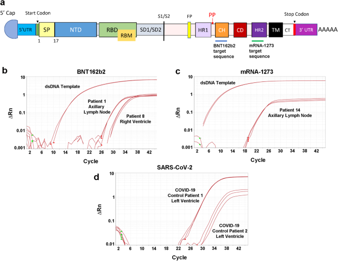
@_HeartofGrace_ - Christie Laura Grace
18/ "Serious adverse complications due to these vaccines are uncommon and may include anaphylactic reactions, myocarditis, pericarditis, myocardial infarction, cerebral sinus thrombosis, stroke, pulmonary embolism, neuropathies, and autoimmune hepatitis" We know, from studies,
@_HeartofGrace_ - Christie Laura Grace
19/ That when you alter the ratio of (+) charged lipids to negative charged (-) modRNA, this would change where it goes in the body. The contamination of DNA plasmids MUST be CHANGING the ZETA potential and overall charge of the LNP, causing it, sometimes, to go to vascular/lung

@_HeartofGrace_ - Christie Laura Grace
20/ Another thing that could be happening, in addition to DNA plasmids changing the zeta potential (charge) is the LNP is breaking down during the freeze/thaw process, and parts can leak out, changing the overall charge as well. Dr. Ko and De, and others proved.


@_HeartofGrace_ - Christie Laura Grace
21/ Negative charges also cause clots (negatively charged liposomes) as shown above. The introduction of the negatively charged DNA plasmids, at a rate of billions per dose, at 100 bp (base pair average, must be shifting zeta. It probably distributes UNEVENLY, which accounts

@_HeartofGrace_ - Christie Laura Grace
22/ for heterogeneity (differences) between batches and vials. Because it is not evenly distributed, there would be batch an vial variation. The FDA needs to investigate immediately, and need to immediately halt distribution. The DNA plasmids are of significant concern.
@_HeartofGrace_ - Christie Laura Grace
23/ https://anandamide.substack.com/p/sequencing-of-bivalent-moderna-and https://anandamide.substack.com/p/south-carolina-senate-hearing

@_HeartofGrace_ - Christie Laura Grace
24/ https://pubmed.ncbi.nlm.nih.gov/36588456/ https://www.nature.com/articles/s42003-021-02441-2